Research Article
IGF-1 LR3 vs PEG-MGF: Growth Factor Comparison
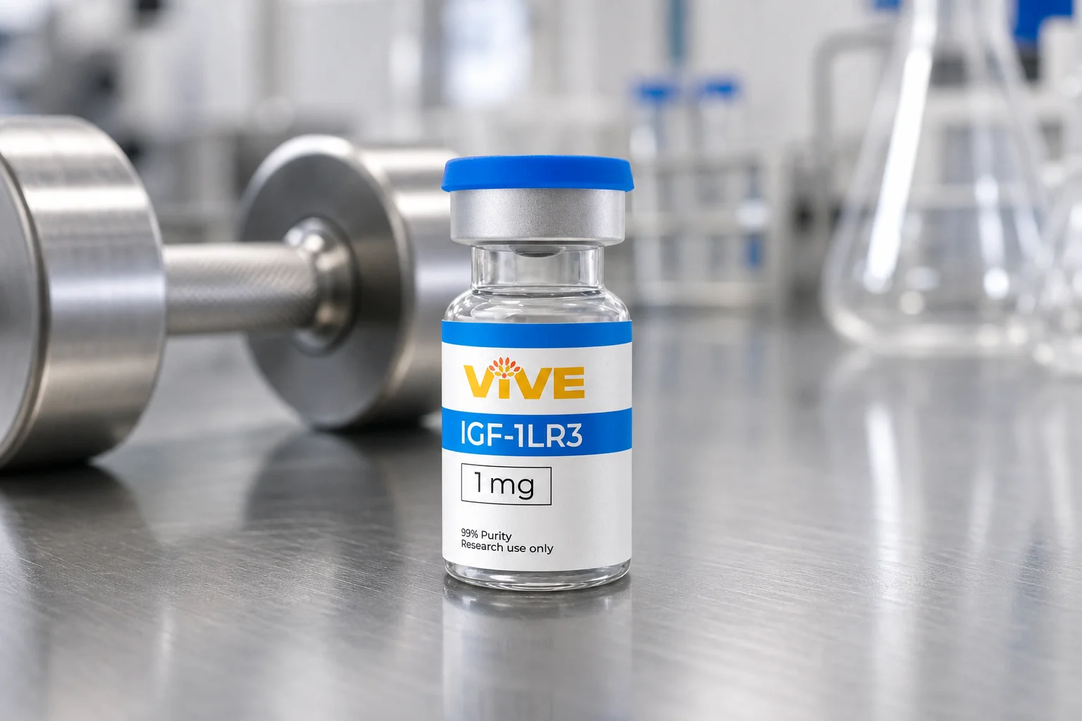
IGF-1 LR3 and PEG-MGF are both splice variant growth factors researchers use to study muscle tissue repair, but they work through different pathways at different timescales. This IGF-1 LR3 vs PEG-MGF comparison breaks down the mechanism of action, half-life, and site of activity researchers weigh before selecting a research-grade IGF-1 LR3 for a given protocol.
By Vive Team
What Is IGF-1 LR3?
IGF-1 LR3 is a long-arginine analog of insulin-like growth factor 1, engineered with a substituted arginine residue at position 3 and a 13-amino acid extension on the N-terminus that resists binding to IGF binding proteins found in serum. In practice, this modification means IGF-1 LR3 circulates longer and produces a more systemic effect than native IGF-1, which is quickly bound and neutralized once released into circulation. Researchers studying satellite cell activation and downstream muscle cell proliferation often select IGF-1 LR3 because its extended half-life allows a single research dose to maintain elevated signaling across a longer observation window than native growth factor would permit. This makes it a common reference point in research comparing systemic versus localized growth factor delivery, and it is one reason IGF-1 LR3 shows up so frequently alongside mechano growth factor in muscle physiology literature.
What Is PEG-MGF?
PEG-MGF is a pegylated form of mechano growth factor (MGF), the splice variant of the IGF-1 gene expressed locally in skeletal muscle after mechanical loading or tissue damage. Where the liver-derived, circulating form of IGF-1 supports long-term systemic growth, mechano growth factor is produced directly inside the muscle fiber itself and acts as a rapid-response signal. Unlike systemic IGF-1 LR3, native mechano growth factor has an extremely short half-life, often described in minutes rather than hours, because it is meant to act at the site of damage and then degrade quickly before it can diffuse into general circulation.
Pegylation extends that window by attaching a polyethylene glycol chain to the peptide backbone, slowing renal clearance and giving researchers a longer usable window for site-specific study designs without eliminating the localized character of the response. Where IGF-1 LR3 is prized for systemic reach, PEG-MGF research protocols instead treat the injection site as the primary zone of activity, since localized action is central to the mechano growth factor pathway and to how researchers interpret its results.
IGF-1 LR3 vs PEG-MGF: Mechanism of Action and Satellite Cell Activation
Both molecules converge on the same downstream target, the satellite cell, but they arrive by different routes. Satellite cells are quiescent muscle stem cells positioned between the sarcolemma and basal lamina of the muscle fiber. When muscle tissue experiences mechanical stress or damage, satellite cell activation is the first checkpoint in the repair cascade: the cell exits quiescence, proliferates, and eventually fuses with existing myofibers or forms new ones entirely.
Mechano growth factor is described in muscle physiology research as an early trigger of this activation at the local site of load or injury, distinguishing it from later-acting, differentiation-focused isoforms of IGF-1 such as IGF-1Ea. IGF-1 LR3, by contrast, works through the classic IGF-1 receptor signaling cascade, activating the PI3K/Akt/mTOR pathway and supporting satellite cells once they have already entered the proliferative and differentiation phase. Researchers frequently frame the pair as sequential rather than redundant: mechano growth factor for initial cell activation, IGF-1 LR3 for sustained downstream signaling and muscle growth support once proliferation is underway.
This sequencing is part of why the two are so often studied side by side rather than as substitutes for one another. A protocol built around one splice variant alone may miss half of the biological picture, since activation and sustained signaling are functionally distinct steps in the same repair pathway.
Half-Life and Site of Action: Systemic vs Localized Research Models
This is where the IGF-1 LR3 vs PEG-MGF distinction becomes a practical research variable rather than an academic one. IGF-1 LR3's resistance to IGF binding proteins gives it a half-life measured in hours, which makes it suitable for study designs that need sustained systemic exposure or repeated-measures protocols where researchers want signaling active across a full observation day. PEG-MGF's extended but still comparatively short half-life keeps its action closer to the injection site, which is useful when the research question centers on localized tissue repair rather than whole-body muscle growth.
Researchers selecting between the two typically start with the question being asked: is the study modeling a systemic growth factor response, or a localized repair response triggered at a specific site? That distinction, more than potency, tends to drive protocol design. Study designs that require repeated sampling across multiple tissue sites, for example, may lean on IGF-1 LR3's longer window to reduce the number of administrations needed, while designs isolating a single site of injury or load may prefer PEG-MGF specifically because its action stays contained rather than becoming systemic.
Reconstitution and Handling Considerations
Both peptides are supplied as lyophilized powder and require reconstitution before use in research settings. Bacteriostatic water is the standard diluent for both, since the benzyl alcohol it contains helps preserve the reconstituted solution across multiple uses. Gentle mixing, swirling rather than shaking, is typically recommended, since vigorous agitation can denature the peptide's tertiary structure and reduce activity in downstream assays. Vive's bacteriostatic water for peptide reconstitution page outlines current concentration and storage guidance used across the catalog.
Once reconstituted, both IGF-1 LR3 and PEG-MGF should be stored refrigerated, generally between 2 and 8 degrees Celsius, and used within the documented stability window for that lot. Repeated freeze-thaw cycles are a common and avoidable source of peptide degradation in lab settings, along with prolonged exposure to light or room-temperature storage after reconstitution. Keeping a written log of reconstitution date, diluent volume, and storage conditions helps researchers correlate any drop-off in observed activity with a handling variable rather than the peptide itself.
Frequently Asked Questions
What is the main difference between IGF-1 LR3 and PEG-MGF? IGF-1 LR3 is a long-acting IGF-1 analog with a multi-hour half-life suited to systemic research exposure, while PEG-MGF is a pegylated mechano growth factor variant with a shorter, more localized window of activity tied to the injection site. Researchers typically select IGF-1 LR3 for systemic study designs and PEG-MGF for site-specific repair models.
Why does mechano growth factor have such a short natural half-life? Native mechano growth factor is expressed locally in muscle tissue in response to mechanical load or damage and is built to act quickly at that site before degrading. This localized, transient expression pattern is part of what distinguishes it from more stable, systemically circulating growth factors like IGF-1 LR3.
Can IGF-1 LR3 and PEG-MGF be studied together in the same protocol? Some research designs pair the two to model both the early activation phase associated with mechano growth factor and the sustained downstream signaling associated with IGF-1 LR3. Study design should follow the specific research question, and researchers should consult current literature and institutional protocols before combining any research peptides.
How should IGF-1 LR3 and PEG-MGF be stored after reconstitution? Both peptides should be reconstituted with bacteriostatic water, kept refrigerated, and used within the documented stability window for that lot. Avoid repeated freeze-thaw cycles and vigorous shaking, both of which can degrade peptide structure and affect the reliability of downstream satellite cell activation assays.
Is either peptide intended for human use outside of research settings? No. IGF-1 LR3 and PEG-MGF are sold strictly for laboratory and research purposes and are not evaluated or approved for human consumption. All handling, storage, and use should follow institutional research protocols and applicable regulations rather than any therapeutic, cosmetic, or clinical use case.
Compare Growth Factor Research Peptides at VivePeptides
Choosing between IGF-1 LR3 and PEG-MGF ultimately comes down to whether a protocol calls for systemic reach or localized, site-specific signaling. Shop research peptides to review current specifications for IGF-1 LR3 and the rest of the growth factor lineup before finalizing your next research order.
Research Use Only
All information in this article is intended for educational and research purposes only. VivePeptides products are not intended for human or veterinary use.