Research Article
7 Best Peptides for Muscle Recovery Research in 2026
Researchers studying musculoskeletal repair have identified a small group of peptides that appear repeatedly across tissue healing models. This guide covers seven of the most rigorously studied compounds in the 2026 muscle recovery research landscape, examining their proposed mechanisms, relevant preclinical data, and how they are being used in laboratory settings.
Research Use Only. All compounds discussed in this article are intended strictly for in-vitro and laboratory research purposes. None are approved for human or veterinary use. This content is for informational and scientific reference only.
Why Peptides Are Central to Muscle Recovery Research
Muscle tissue repair is a multi-phase biological process. After acute injury or intense mechanical stress, satellite cells activate, inflammatory cascades are triggered, and the extracellular matrix undergoes significant remodeling. Each of these phases is regulated by signaling molecules, many of which are peptides.
Research interest in exogenous peptides for musculoskeletal recovery stems from their ability to interact with specific receptors and signaling pathways involved in these repair processes. Unlike broad-spectrum compounds, peptides tend to operate with a degree of mechanistic specificity that makes them valuable tools in controlled research environments.
A 2024 systematic review published in Orthopaedic Journal of Sports Medicine analyzed 36 studies and found that peptides targeting angiogenesis, cellular migration, and growth factor expression consistently showed measurable effects on tissue repair endpoints in preclinical models. That body of evidence is driving continued investigation into which peptide structures and combinations most reliably support skeletal muscle recovery under laboratory conditions.
The following seven compounds represent the current frontline of this research.

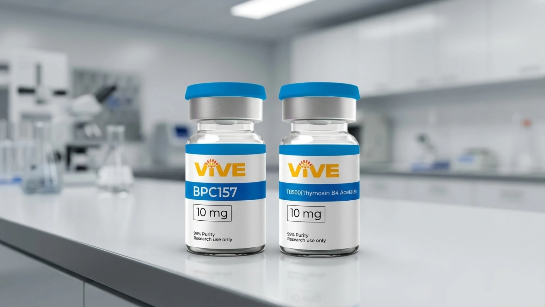
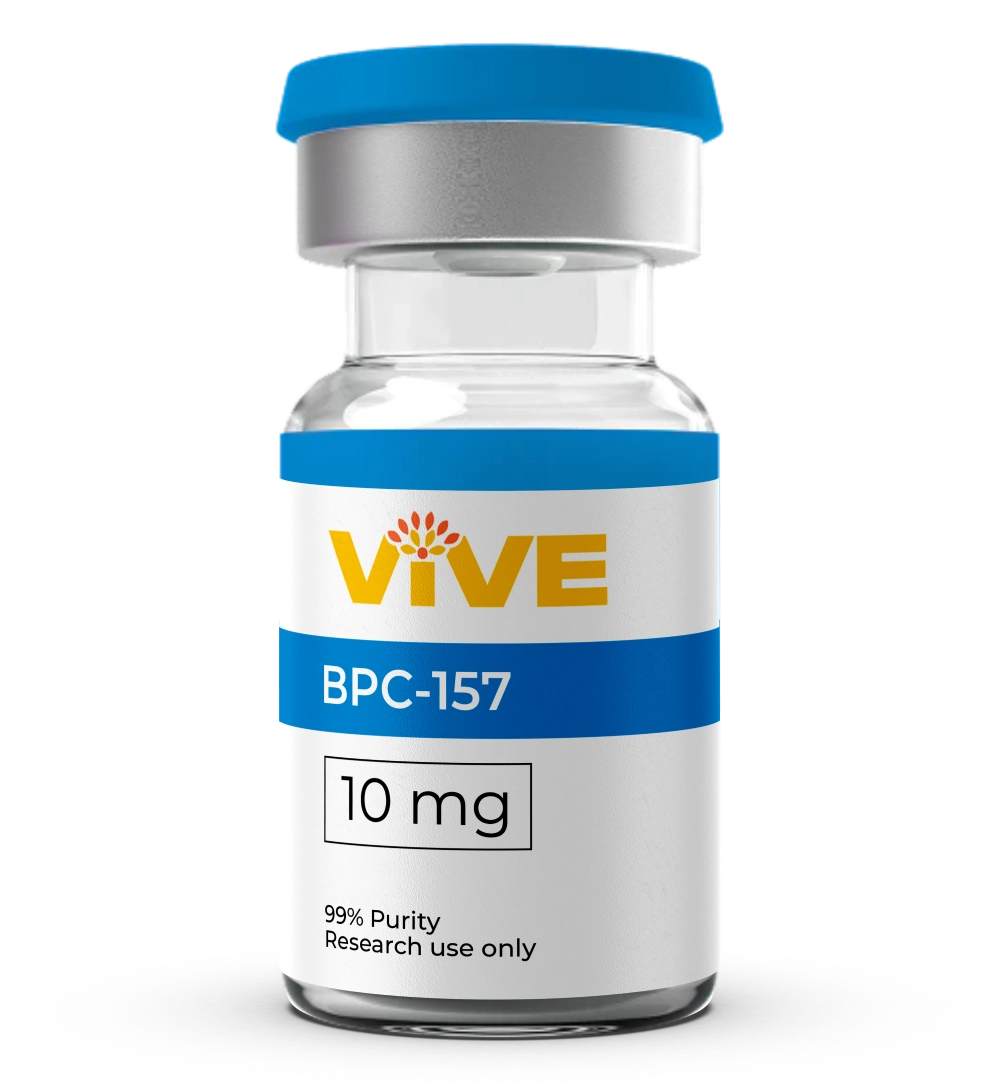
1. BPC-157 (Body Protection Compound 157)
Top Researched
BPC-157 is a synthetic 15-amino acid peptide derived from a protective protein found in gastric juice. It is among the most extensively studied peptides in the musculoskeletal literature, with research spanning muscle, tendon, ligament, and bone injury models.
Proposed Mechanism
BPC-157 appears to upregulate vascular endothelial growth factor (VEGF) and epidermal growth factor (EGF) expression at injury sites, accelerating angiogenesis and collagen type I deposition. At the cellular level, research by Dr. Predrag Sikiric and colleagues at the University of Zagreb has documented enhanced paxillin and vinculin expression, proteins involved in focal adhesion signaling that support muscle cell migration and spreading.
A study published in the Journal of Physiology examined BPC-157 in skeletal muscle crush models and reported accelerated satellite cell activation and faster restoration of contractile function compared to control groups. Subsequent work demonstrated that these effects persist even when systemic corticosteroids are present, a finding that distinguishes BPC-157 from several other repair-signaling compounds.
Research Relevance in 2026
The 2025 systematic review by Vasireddi, Hahamyan, Salata, and colleagues, published in Orthopaedic Journal of Sports Medicine, analyzed 35 preclinical and one clinical study and concluded that BPC-157 consistently improved outcomes in muscle tear, tendon rupture, and ligament injury models. Satellite cell activation and VEGF-driven angiogenesis were the most frequently reported mechanisms.
For researchers focused on muscle repair signaling, BPC-157 remains the compound with the broadest preclinical dataset.
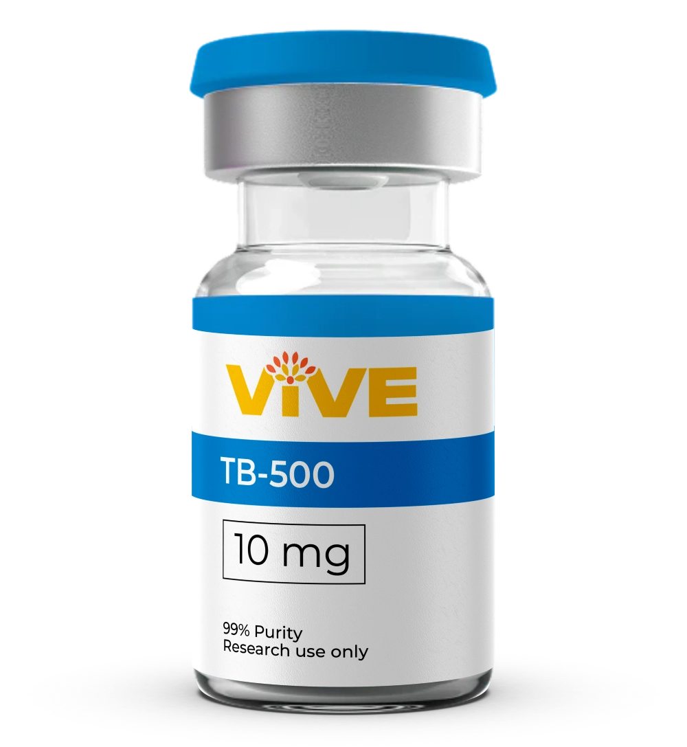
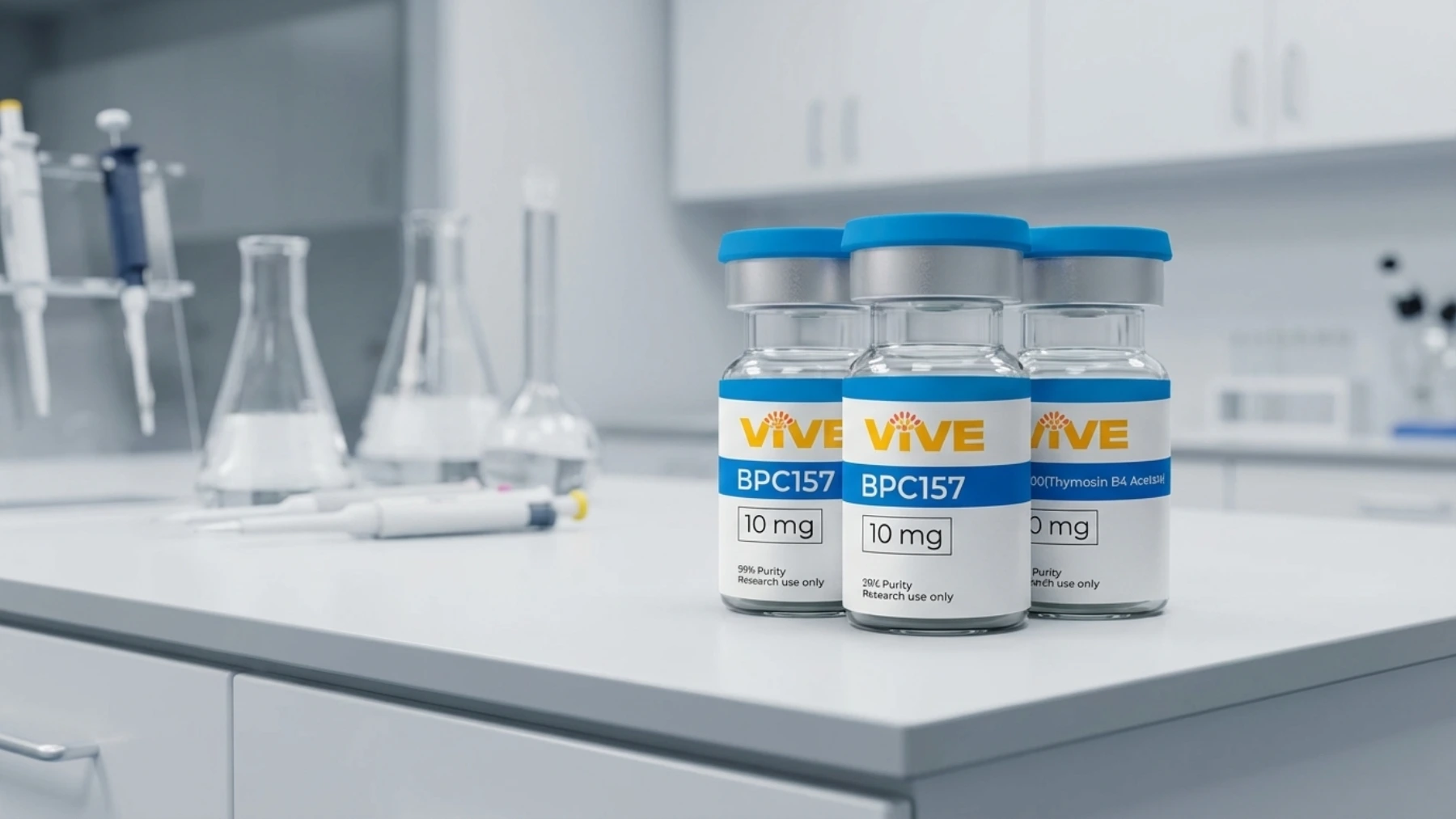
2. TB-500 (Thymosin Beta-4 Acetate)
Repair Signaling
TB-500 is a 43-amino acid synthetic peptide derived from thymosin beta-4, a naturally occurring protein with known roles in actin regulation and cellular development. It operates through a fundamentally different mechanism than BPC-157, making it a complementary compound in multi-peptide recovery research protocols.
Proposed Mechanism
TB-500's primary biological activity involves binding to G-actin monomers. By sequestering actin monomers, the peptide modulates actin polymerization, which directly influences cellular motility. This mechanism promotes migration of endothelial cells, myocardial cells, and tissue progenitor cells toward injury sites.
Research published in the Journal of Cell Science demonstrated that thymosin beta-4 upregulates VEGF expression through pathways distinct from those activated by BPC-157, meaning both compounds can drive angiogenesis simultaneously without redundant pathway competition.
In experimental muscle injury models, TB-500 administration has been observed to accelerate regeneration of damaged muscle fibers, reduce inflammatory infiltration, and increase flexibility in connective tissue adjacent to the repair site. A 2021 study published in PMC (NCBI) examining thymosin beta-4's role in regenerative tissue contexts noted that the peptide's actin-binding specificity gives it a mechanistic profile unlike most growth-factor-based compounds.
Why Researchers Pair TB-500 with BPC-157
The two compounds act at different stages of the repair cascade. BPC-157 primarily drives early growth factor expression and satellite cell recruitment, while TB-500 facilitates cellular migration toward the site of injury. In combination research protocols, this complementarity has made them a frequently studied pairing.
3. IGF-1 LR3 (Insulin-Like Growth Factor 1 Long R3)
Hypertrophy Research
IGF-1 LR3 is a structurally modified analog of native IGF-1. The "LR3" designation refers to two specific modifications: an arginine-to-glutamic acid substitution at position 3 and a 13-amino acid N-terminal extension. These changes reduce binding affinity to IGF-binding proteins (IGFBPs) by more than 100-fold, extending the peptide's half-life from minutes (native IGF-1) to approximately 20 to 30 hours.
Proposed Mechanism
IGF-1 LR3 interacts with IGF-1 receptors expressed on muscle satellite cells, driving two parallel signaling cascades. The PI3K/Akt/mTOR pathway promotes protein synthesis, while the MAPK/ERK pathway stimulates satellite cell proliferation and differentiation. In muscle repair research, satellite cell activation is a central outcome variable, as these cells are the primary regenerative population responsible for rebuilding damaged myofibers.
Research published in the American Journal of Physiology and reviewed in PMC (PMC4665094) documented that sustained IGF-1 receptor activation through extended-half-life analogs produced measurable increases in myonuclear addition, a process that theoretically expands a muscle fiber's capacity for ongoing repair and adaptation.
Extended Activity Window
The IGFBP-resistant structure of IGF-1 LR3 means the peptide remains biologically available for a significantly longer period than native IGF-1. Researchers studying muscle hypertrophy signaling use this extended activity window to examine how sustained IGF-1 receptor occupancy affects satellite cell kinetics and downstream anabolic pathway regulation.
Most available evidence comes from rodent and in vitro models. Human clinical data on IGF-1 LR3 specifically remains limited.
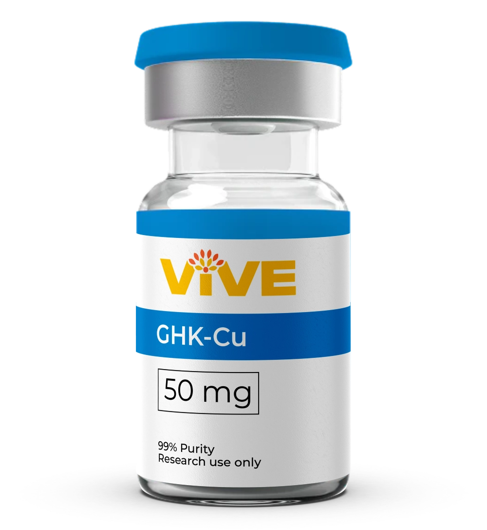
4. GHK-Cu (Copper Peptide)
Matrix Remodeling
GHK-Cu is a naturally occurring tripeptide (glycine-histidine-lysine) complexed with copper ions. It is among the most structurally characterized peptides in the literature, with research dating back to the 1970s when Dr. Loren Pickart first identified its presence in human plasma and its ability to stimulate tissue repair responses.
Proposed Mechanism
At the extracellular matrix level, GHK-Cu simultaneously stimulates synthesis of collagen types I and III, glycosaminoglycans, elastin, and proteoglycan decorin. It also upregulates matrix metalloproteinase activity to degrade and remove damaged collagen, enabling proper matrix remodeling rather than fibrotic scar deposition. This dual action, building new matrix while clearing damaged material, gives GHK-Cu a unique profile in tissue repair research.
A 2018 review published in the International Journal of Molecular Sciences (PMC6073405) by Dr. Loren Pickart and colleagues documented that GHK modulates expression of more than 4,000 human genes, including pathways involved in DNA repair, anti-inflammatory signaling, and mitochondrial function.
Relevance to Muscle Recovery Research
In the context of muscle repair, GHK-Cu's significance lies in its action on connective tissue, tendons, and the fascial structures that surround and support muscle groups. Recovery from intense training or injury is not limited to muscle fiber regeneration; the surrounding matrix must also be remodeled appropriately. Research involving GHK-Cu in wound healing models has demonstrated accelerated collagen deposition and improved mechanical properties of repaired tissue.
Plasma levels of GHK decline with age, from approximately 200 ng/mL at age 20 to roughly 80 ng/mL by age 60, a reduction that researchers have proposed as a contributing factor in age-related declines in tissue repair capacity.
5. MOTS-c (Mitochondrial Open Reading Frame of the 12S rRNA-c)
Metabolic Research
MOTS-c is a 16-amino acid peptide encoded within the 12S ribosomal RNA region of the mitochondrial genome, making it one of the few known mitochondrial-derived peptides with documented effects on skeletal muscle metabolism. Its discovery in 2015 by Dr. Changhan David Lee and colleagues at the University of Southern California opened a new line of inquiry into how mitochondria communicate with the nucleus and systemic tissues.
Proposed Mechanism
MOTS-c is transcribed in response to metabolic stress and exercise. Once secreted from mitochondria, it translocates to the nucleus and regulates the expression of stress-adaptation genes, primarily through the Folate-AICAR-AMPK pathway. AMPK activation has downstream effects on mitochondrial biogenesis, glucose uptake, and fatty acid oxidation, all relevant variables in exercise physiology research.
A study published in Nature Communications (2021) found that endogenous MOTS-c levels in skeletal muscle increased approximately 11.9-fold following exercise, with circulating levels rising 1.6-fold during and 1.5-fold after exercise before returning to baseline within four hours. This exercise-responsive expression pattern positions MOTS-c as a potential mediator of the metabolic adaptations associated with training.
Separately, research published in the American Journal of Physiology-Endocrinology and Metabolism demonstrated that MOTS-c reduces myostatin expression and muscle atrophy signaling, suggesting a role not only in energy metabolism but in the preservation of muscle mass under catabolic conditions.
Why MOTS-c Appears in Muscle Recovery Research
Muscle recovery is not purely a structural repair process. Energy substrate availability, mitochondrial efficiency, and atrophy resistance all influence how quickly and how completely muscle tissue recovers from stress. MOTS-c's ability to act on these metabolic variables gives it a distinct research profile compared to the structural repair peptides discussed above.
6. CJC-1295 + Ipamorelin Blend
GH Axis Research
The combination of CJC-1295 (a GHRH analog) and Ipamorelin (a selective GH secretagogue) represents a different research angle: modulating growth hormone axis signaling rather than acting directly on repair pathways.
Proposed Mechanism
CJC-1295 is a 30-amino acid analog of growth hormone-releasing hormone (GHRH). It binds to the GHRH receptor on pituitary somatotroph cells, stimulating pulsatile GH release. Ipamorelin operates through the ghrelin receptor (GHS-R1a), producing a GH pulse without the significant cortisol or prolactin elevation seen with older secretagogues like GHRP-2.
When combined, the two compounds produce a synergistic GH pulse substantially larger than either compound alone, according to endocrinology research examining GHRH/ghrelin receptor co-activation.
Connection to Muscle Recovery
Growth hormone drives downstream IGF-1 production in the liver, which then acts on muscle satellite cells and connective tissue. In preclinical models, GH axis activation has been associated with improved nitrogen retention, accelerated protein synthesis, and enhanced recovery from exercise-induced muscle damage. Researchers studying sleep quality and nocturnal GH secretion patterns also use CJC-1295 + Ipamorelin protocols to investigate how GH pulse timing affects recovery endpoints.
Importantly, Ipamorelin's selectivity for the GH axis without significant cortisol effects makes this pairing a commonly modeled protocol in controlled study designs.
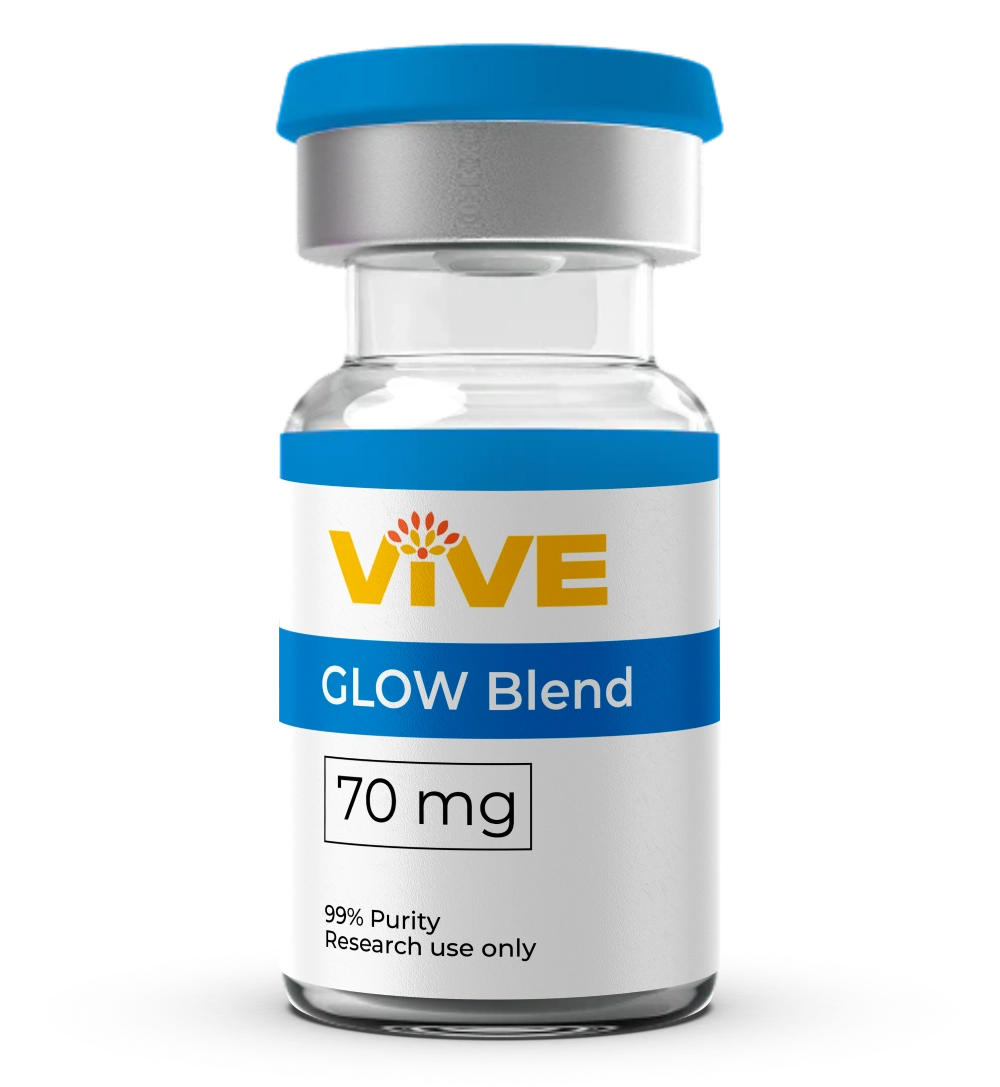
7. Glow Blend (BPC-157 + TB-500 + GHK-Cu)
Multi-Pathway Stack
The Glow Blend combines three of the compounds from this list, BPC-157, TB-500, and GHK-Cu, into a single research compound. The rationale for multi-peptide combinations in research is mechanistic coverage: different peptides act at different stages and pathways of the repair cascade.
Why Multi-Peptide Research Combinations Are Being Studied
- BPC-157 drives early growth factor expression, satellite cell recruitment, and angiogenesis
- TB-500 facilitates cellular migration to injury sites via actin regulation
- GHK-Cu supports extracellular matrix remodeling and connective tissue integrity
Each operates through distinct receptors and pathways, minimizing competitive interference while potentially producing additive effects across the full repair process. Research published in Orthopaedic journals in 2024 and 2025 noted that multi-compound protocols are increasingly common in preclinical tissue repair studies precisely because isolated peptide effects may not reflect the full complexity of in vivo repair signaling.
For researchers studying comprehensive recovery models rather than isolating single mechanisms, combination compounds offer a practical and methodologically coherent approach.
Side-by-Side Research Comparison
Peptide | Primary Research Target | Key Mechanism | Evidence Base
---|---|---|---
BPC-157 | Muscle, tendon, ligament | VEGF upregulation, satellite cell activation | 35+ preclinical studies (2025 systematic review)
TB-500 | Muscle fiber, connective tissue | G-actin sequestration, cellular migration | Preclinical, in vitro
IGF-1 LR3 | Satellite cell kinetics, hypertrophy | PI3K/Akt/mTOR + MAPK/ERK activation | Preclinical, in vitro
GHK-Cu | Extracellular matrix, connective tissue | Collagen synthesis + MMP-mediated remodeling | In vitro, wound models, 4,000+ gene interactions
MOTS-c | Muscle metabolism, atrophy resistance | AMPK activation, myostatin reduction | Preclinical, one human exercise study
CJC-1295 + Ipamorelin | GH axis, systemic recovery | Pulsatile GH release, downstream IGF-1 | Preclinical, clinical GH axis studies
Glow Blend | Multi-pathway muscle and tissue | BPC-157 + TB-500 + GHK-Cu combined | Component-level evidence; stacking rationale
Choosing the Right Compound for Your Research Protocol
The appropriate peptide or combination depends on the specific recovery pathway your research is targeting.
Structural Repair Focused
Researchers targeting muscle fiber regeneration, satellite cell activation, and angiogenesis at the injury site typically center protocols around BPC-157, TB-500, or both in combination.
Matrix Remodeling Focused
Studies examining connective tissue integrity, collagen quality, and fascial recovery benefit from GHK-Cu's dual synthesis and degradation activity on the extracellular matrix.
Metabolic Recovery Focused
Protocols investigating mitochondrial efficiency, atrophy signaling, and metabolic adaptation to training stress find MOTS-c's AMPK-mediated mechanisms most directly relevant.
Multi-Pathway Coverage
Research designs requiring broad mechanistic coverage across repair stages use combination compounds like the Glow Blend or layer CJC-1295 + Ipamorelin for GH axis support.
All seven compounds discussed here are available in research-grade form, verified at 99%+ purity by independent third-party HPLC and mass spectrometry analysis. Explore the full catalog at VivePeptides Shop All Peptides.
Purity Standards: Every compound at VivePeptides ships with a batch-specific Certificate of Analysis (COA) confirming identity, purity, and third-party testing results. Research integrity starts with compound quality.
Frequently Asked Questions
What peptides are most studied for muscle recovery research?
BPC-157 and TB-500 have the largest preclinical literature bases specifically related to musculoskeletal repair. A 2025 systematic review in Orthopaedic Journal of Sports Medicine identified BPC-157 as the most frequently studied peptide in orthopaedic soft tissue injury models, with 36 studies analyzed. MOTS-c and IGF-1 LR3 are gaining traction in research focused on metabolic and hypertrophy dimensions of muscle recovery.
How do BPC-157 and TB-500 differ in their mechanisms?
BPC-157 primarily drives growth factor expression and satellite cell activation at the injury site, while TB-500 acts through actin-binding to promote cellular migration toward the injury. The two compounds operate through separate receptor systems and are considered mechanistically complementary in research protocols that aim to cover multiple phases of the repair cascade.
What is MOTS-c and why is it relevant to muscle research?
MOTS-c is a mitochondrial-derived peptide that activates the AMPK signaling pathway in response to metabolic stress. Research published in Nature Communications found that skeletal muscle MOTS-c levels increase nearly 12-fold following exercise. Its ability to reduce myostatin expression and modulate mitochondrial biogenesis makes it relevant to both endurance metabolism and muscle preservation research.
What does GHK-Cu do in tissue repair research?
GHK-Cu stimulates collagen type I and III synthesis while simultaneously upregulating matrix metalloproteinases to clear damaged collagen. This dual activity supports proper extracellular matrix remodeling, as opposed to disorganized fibrotic repair. Research has also identified GHK-Cu as a modulator of more than 4,000 human genes, including those involved in DNA repair and anti-inflammatory signaling.
Where can I source research-grade peptides for these studies?
Research-grade compounds require verified purity, batch-specific documentation, and proper handling certification. VivePeptides provides all seven compounds discussed in this article, each verified at 99%+ purity by independent U.S. laboratories with HPLC and mass spectrometry analysis. View available compounds at vivepeptides.com/shop.
Research Use Only
All information in this article is intended for educational and research purposes only. VivePeptides products are not intended for human or veterinary use.