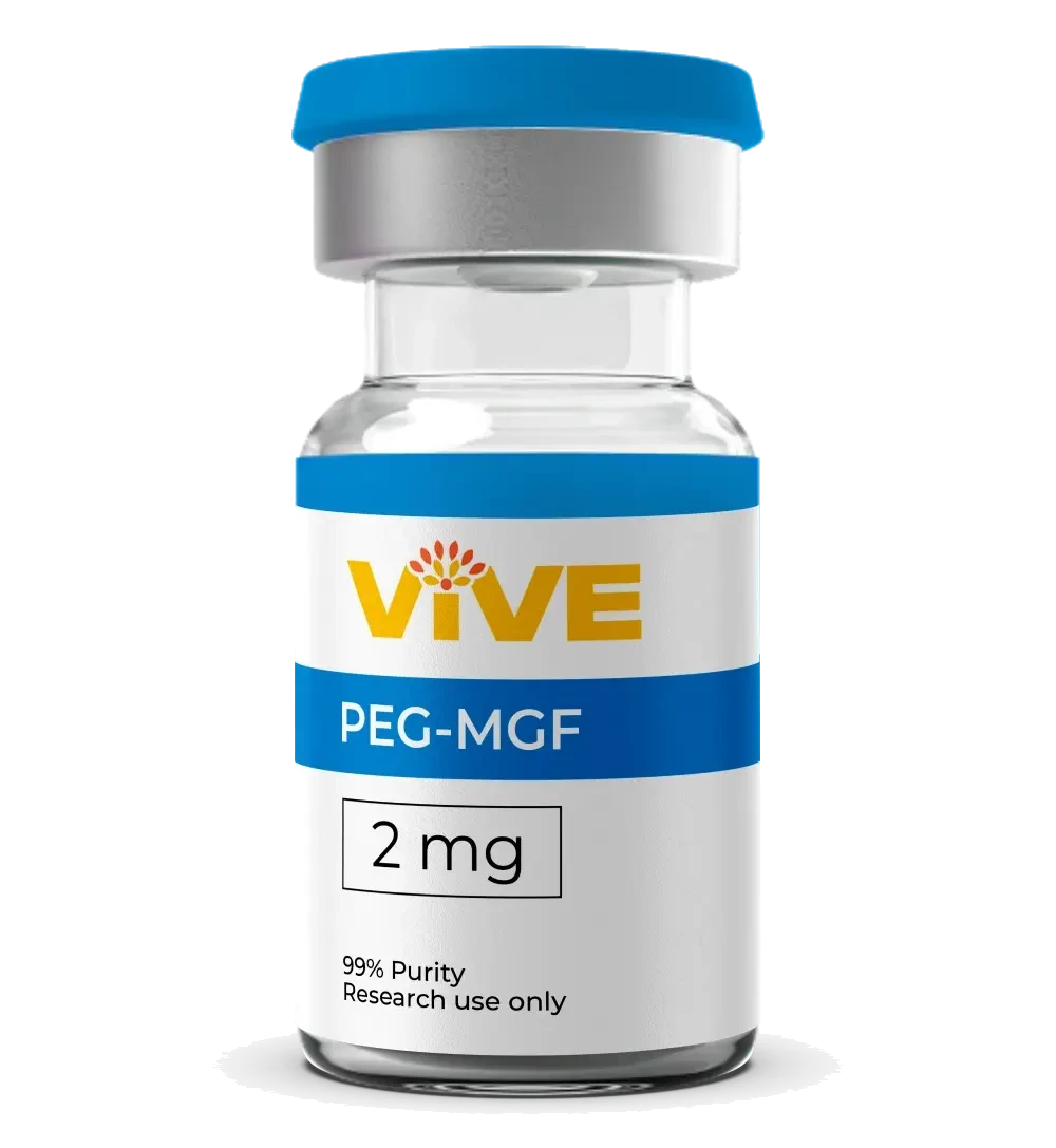
PEG-MGF
- Size
- 2mg
Specifications
PEG-MGF Technical Profile
OVERVIEW
What Is PEG-MGF Peptide?
PEG-MGF (Pegylated Mechano Growth Factor) is a synthetic peptide analog derived from the C-terminal E-domain of IGF-1Ec, the mechano-sensitive splice variant of insulin-like growth factor 1. In natural physiology, MGF (mechano growth factor) is expressed locally in skeletal muscle and other tissues in response to mechanical loading and microtrauma. The pegylated form, PEG-MGF peptide, incorporates a polyethylene glycol (PEG) moiety that significantly extends the compound's serum half-life and stability compared to native MGF, making it a preferred analog for controlled laboratory and animal-model research protocols.
As a research peptide, PEG-MGF has attracted scientific attention for its proposed role in satellite cell activation and local muscle tissue biology. Unlike the parent IGF-1 molecule, which circulates systemically and signals through the canonical IGF-1 receptor with broad anabolic effects, the MGF E-domain is understood in preclinical literature to act through a distinct, locally mediated mechanism. VivePeptides supplies PEG-MGF 2mg vials at research-grade purity for laboratory investigation. This compound is supplied for research use only and is not approved for human or veterinary use.
- 01
Pegylated IGF-1Ec Analog
Synthetic peptide derived from the C-terminal E-domain of the IGF-1Ec (mechano growth factor) splice variant, with PEG modification for extended stability
- 02
≥99% HPLC Purity
Every batch verified via high-performance liquid chromatography to confirm identity, sequence integrity, and purity at or above 99%
- 03
USA Tested and Verified
Third-party analytical testing performed at USA-based laboratories, with documentation available for every production lot
RESEARCH
PEG-MGF Peptide Mechanism of Action in Research
The research focus on PEG-MGF centers on its proposed role as a locally acting signal in skeletal muscle satellite cell biology. Satellite cells are quiescent myogenic progenitor cells that reside between the sarcolemma and the basal lamina of muscle fibers. Preclinical literature suggests that the MGF E-domain peptide acts on these satellite cells through a receptor or binding interaction distinct from the classic IGF-1R pathway, stimulating their proliferation before they differentiate and fuse with existing muscle fibers. Goldspink G. and colleagues were instrumental in characterizing this isoform and its distinct functional profile relative to systemic IGF-1.
Pegylation modifies the pharmacokinetic profile of the peptide substantially. Native MGF is degraded rapidly in serum, with a half-life measured in minutes under physiological conditions. The PEG modification confers resistance to proteolytic degradation, prolonging the window during which the peptide remains detectable in preclinical assay systems. This extended stability is one of the primary reasons researchers select PEG-MGF peptide over native MGF when designing time-course studies in animal models.
Satellite Cell Activation Research
Preclinical studies indicate that the MGF E-domain peptide may stimulate the proliferation of skeletal muscle satellite cells, the resident stem-cell population responsible for postnatal muscle repair and adaptation. Hill and Goldspink (2003) reported that MGF promotes satellite cell activation in animal models, an effect proposed to precede the differentiation phase that follows muscle fiber damage. This line of research has positioned PEG-MGF as a tool for investigating the early cellular events in muscle tissue biology.
Distinct Signaling From Canonical IGF-1 Pathway
A key finding in the MGF peptide literature is that the C-terminal E-domain does not appear to signal through the standard IGF-1 receptor (IGF-1R) in the same manner as mature IGF-1. Yang and Goldspink (2002) published work demonstrating that MGF and IGF-1 Ea exert different effects on muscle cells in culture, suggesting receptor or co-receptor specificity that is still being characterized in preclinical research. This mechanistic distinction is central to the scientific rationale for studying PEG-MGF separately from other IGF-1 isoforms.
Mechano-Sensitive Expression and Loading Models
Research by Hameed et al. documented that MGF mRNA expression is upregulated in skeletal muscle following resistance-type loading protocols in human and animal subjects, making it one of the few peptides with a well-characterized mechanical stimulus for expression. Laboratory models studying the IGF-1Ec splice variant use resistance loading or stretch protocols to trigger endogenous MGF expression, and exogenous PEG-MGF peptide has been applied in parallel studies to isolate the E-domain's specific contribution to satellite cell dynamics.
PEG Modification and Extended Half-Life
The polyethylene glycol chain attached to MGF substantially alters its degradation kinetics in preclinical assay systems. While native MGF is cleared rapidly, PEG-MGF demonstrates a prolonged presence in circulation in animal models, enabling researchers to study time-dependent biological responses that would be impossible to capture with the unmodified peptide. This pharmacokinetic advantage is the primary structural rationale for the pegylated analog in controlled research settings.
COMPARISON
PEG-MGF vs Native MGF: Research Peptide Comparison
PEG-MGF
PEG-MGF and native MGF share the same core E-domain peptide sequence derived from the IGF-1Ec isoform, but differ substantially in their stability, duration of action in preclinical assay systems, and practical utility in controlled laboratory protocols. Understanding these differences is important for researchers selecting the appropriate analog for their experimental design.
Native MGF
IGF-1 LR3 is frequently referenced alongside MGF peptide analogs in the research literature, as it is also derived from the IGF-1 family. However, IGF-1 LR3 is a full-length IGF-1 analog with an arginine substitution at position 3, acts systemically through the IGF-1R, and does not replicate the locally acting, satellite-cell-specific biology attributed to the MGF E-domain. VivePeptides offers both PEG-MGF 2mg and related research peptides at ≥99% HPLC purity from our USA-tested catalog.
| Feature | PEG-MGF | Native MGF | IGF-1 LR3 (reference) |
|---|---|---|---|
| Origin | Synthetic pegylated C-terminal E-domain of IGF-1Ec | C-terminal E-domain of IGF-1Ec splice variant | Full-length IGF-1 analog with Arg3 substitution |
| Modification | PEG chain attached for extended stability | No modification, native sequence | Arg3 substitution plus 13-aa N-terminal extension |
| Serum Half-Life (preclinical) | Extended (hours) due to PEG moiety | Short (minutes), rapidly degraded | Extended (hours to days) |
| Primary Receptor | Distinct from canonical IGF-1R (under investigation) | Distinct from canonical IGF-1R (under investigation) | IGF-1R (canonical), low IGFBP affinity |
| Research Focus | Satellite cell activation, local muscle tissue biology | Satellite cell activation, mechano-sensitive expression | Systemic anabolic signaling, cell proliferation models |
| Action Profile | Locally acting analog with extended window | Locally acting, rapid clearance | Systemic, broad IGF-1R-mediated activity |
| Practical Lab Use | Preferred over native MGF for time-course studies | Used in acute expression models | Used in systemic IGF-1 signaling studies |
| Vial Size (VivePeptides) | 2mg | Not offered (unstable form) | 1mg / 2mg |
RESEARCH STUDIES
MGF Peptide Research Applications and Published Studies
The preclinical literature on mechano growth factor and PEG-MGF spans foundational molecular characterization through applied animal-model studies. The research below represents the primary published investigations that established the scientific basis for studying this peptide analog. All findings are preclinical and are presented for informational purposes in the context of research use only.
Goldspink: MGF Characterization and Isoform Biology
Professor Geoffrey Goldspink and colleagues were the first to characterize MGF as a distinct splice variant of IGF-1, identifying the unique C-terminal E-domain that differentiates it from IGF-1 Ea and IGF-1 Eb. This foundational work established that the IGF-1Ec isoform is preferentially expressed in mechanically loaded muscle tissue rather than liver, a distribution pattern that distinguished it from the systemic IGF-1 axis and provided the scientific rationale for subsequent research into its local satellite-cell-activating properties. This body of work defines the conceptual framework for all PEG-MGF peptide research.
Yang and Goldspink (2002): Differential Effects on Muscle Cells
Yang and Goldspink (2002) published a study in FEBS Letters demonstrating that MGF and the IGF-1 Ea isoform produce distinct responses when applied to muscle cells in culture. The data indicated that MGF stimulated an increase in myoblast number while IGF-1 Ea promoted differentiation, suggesting that MGF functions primarily to expand the satellite cell pool rather than drive terminal differentiation. This study is one of the most frequently cited pieces of evidence for the mechanistically distinct role of the MGF E-domain in skeletal muscle research.
Hill and Goldspink (2003): Satellite Cell Activation
Hill and Goldspink (2003) reported observations of MGF in satellite cell activation using animal models of muscle loading and injury. Their findings, published in the Journal of Endocrinology, indicated that MGF expression preceded the upregulation of IGF-1 Ea following muscle damage, consistent with a model in which MGF initiates the satellite cell proliferation phase while other IGF-1 isoforms sustain subsequent growth and differentiation. This temporal sequence has informed the design of preclinical studies using PEG-MGF peptide to isolate the early-activation stage.
Hameed et al.: MGF Expression with Resistance Loading
Hameed and colleagues documented MGF mRNA expression in human and animal skeletal muscle following resistance loading protocols, providing evidence that mechanical strain is a primary driver of local IGF-1Ec expression. Their work corroborated the mechano-sensitive nature of MGF described in earlier preclinical models and supported the use of exogenous PEG-MGF peptide as a research tool to study the downstream cellular consequences of this mechanically induced signal. These findings established a translational rationale for animal-model work with the pegylated analog.
QUALITY ASSURANCE
Quality and Testing Standards for PEG-MGF 2mg
VivePeptides maintains rigorous quality control for every batch of PEG-MGF peptide. Our manufacturing and testing standards are designed to ensure that research laboratories receive a consistent, well-characterized compound suitable for controlled preclinical investigation. All PEG-MGF 2mg vials are supplied as white lyophilized powder, desiccated and sealed for long-term stability.
HPLC and Mass Spectrometry Verification
Every batch of VivePeptides PEG-MGF undergoes high-performance liquid chromatography (HPLC) analysis to confirm purity at or above 99%, along with mass spectrometry to verify the correct molecular identity and integrity of the pegylated peptide sequence. These two complementary analytical methods ensure both purity and structural authenticity of the final research compound.
Third-Party USA Laboratory Testing
All PEG-MGF peptide supplied by VivePeptides is independently tested through third-party analytical laboratories based in the USA. Independent verification removes manufacturer bias from the purity and identity claims. Testing documentation is available for every production lot upon request by qualified researchers.
≥99% Purity Standard
VivePeptides PEG-MGF consistently meets or exceeds ≥99% purity as determined by HPLC analysis. This standard ensures that preclinical experiments use a compound with minimal impurities that could confound results, supporting data reproducibility and integrity across research protocols.
Storage and Handling Specifications
PEG-MGF 2mg vials are lyophilized to white powder and should be stored at -20°C in a desiccated environment, protected from light and humidity. For reconstitution, bacteriostatic water or sterile water is recommended. Once reconstituted, the peptide solution should be stored at 2 to 8°C and used promptly to preserve activity in research applications.
FAQ
Frequently Asked Questions About PEG-MGF
What is PEG-MGF used for in research?
What is the difference between PEG-MGF and native MGF?
What purity is VivePeptides PEG-MGF?
How should PEG-MGF 2mg be stored?
How does PEG-MGF relate to IGF-1 in research literature?
Add To Research Cart
PEG-MGF
99%+ purity · USA lab tested · secure shipping
You May Also Research